ff

Tuesday, March 7, 2017

Blood Tests and Biomarkers

Blood Tests and Biomarkers

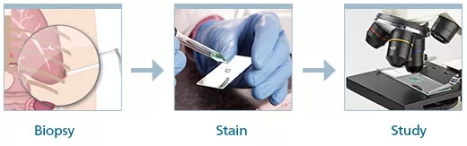
While imaging tests and tissue biopsies are the most common methods for diagnosing mesothelioma, blood tests can also help doctors identify the disease. These blood tests look for certain indicators of disease, known as biomarkers, in a patient’s blood.

MESOMARK Assay


Dr. Snehal Smart of Asbestos.com talks about the MESOMARK assay.
The MESOMARK assay measures the quantity of SMRP in a patient's blood serum. An assay is a test performed in a lab. Abnormally high SMRP levels may indicate the presence of mesothelioma, sometimes years before an asbestos-related cancer diagnosis is made. However, because some types of mesothelioma tumors do not release high levels of SMRP (such as sarcomatoid mesothelioma), the FDA recommends that doctors combine the MESOMARK assay with other tests to ensure an accurate diagnosis. Other diseases that can elevate SMRP levels include pancreatic cancer, ovarian cancer, lung cancer and pulmonary carcinomas.
Fast Fact: In a study of 1,086 blood samples, 99 percent of the healthy specimens displayed SMRP levels at or below the reference value used to indicate the presence of an asbestos-related cancer.
MESOMARK can also help doctors track the progression of a mesothelioma tumor. In most cases, SMRP levels increase as the disease progresses. In one study, 10 out of 18 patients whose imaging scans indicated progressive disease also displayed an SMRP level increase of at least 30 percent on a follow-up MESOMARK assay.

Free Mesothelioma Guide

Free information, books, wristbands and more for patients and caregivers.
Get Your Free Guide
Free Mesothelioma Packet

The MESOMARK Assay Procedure

The MESOMARK assay is completed in two steps. First, the doctor takes two small blood samples from the patient's vein. Each sample contains about two teaspoons of blood. The doctor then sends the samples to a laboratory for official analysis. The patient is free to go home after the procedure, and is generally not at risk for any significant side effects.
At the laboratory, technicians add several antibodies to the blood samples that bind to the protein fragments. They then stir the mixture and place it in a laboratory instrument called a plate reader. Computer software analyzes the contents of the blood sample and provides a measurement of the amount of SMRP-bound antibodies present.

Percent of patients who had elevated serum SMRP levels

Elevated Serum SMRP levels bar graph
SMRP levels at or above a certain reference point may suggest the presence mesothelioma or another malignant pleural disease. Elevated SMRP levels can also be used to identify asbestos-exposed individuals who do not yet have the illness. This implies that blood tests could someday serve as a screening tool for patients at high risk for developing asbestos-related cancer in the future, but most studies suggest the results are not yet reliable enough.
In one study of 40 subjects with a history of asbestos exposure, seven individuals displayed elevated SMRP levels. Within five years of the blood test, three of these patients were diagnosed with the disease. None of the 33 patients with normal SMRP levels developed mesothelioma in the next eight years.

N-ERC/Mesothelin Test

Created in Japan, this blood test detects a form of mesothelin known as N-ERC/mesothelin. This test is similar to MESOMARK in its design, but uses a specially designed enzyme test to increase accuracy.
The N-ERC/mesothelin test is 95 percent effective at detecting mesothelioma, but only 76 percent effective at ruling it out.
Even though this test is more accurate than MESOMARK, it cannot serve as a single test to diagnosed mesothelioma because mesothelin is produced by other cancers than mesothelioma.
The N-ERC test is more accurate at detecting epithelial mesothelioma than sarcomatoid. This is because epithelial mesothelioma produces more mesothelin than the sarcomatoid cell type.

Fibulin-3 Test

Fibulin-3 is a protein overexpressed by mesothelioma and found in pleural fluid and blood.
Mesothelioma specialist Dr. Harvey Pass helped pioneer the fibulin-3 test for mesothelioma. In 2012, he co-authored a study that found fibulin-3 96.7 percent effective at detecting mesothelioma and 95.5 percent effective at ruling it out.
Pass also reported that when used to detect pleural mesothelioma among asbestos-exposed persons, the fibulin test had 100 percent sensitivity and 94.1 percent specificity. However, when the test was used on archived blood serum samples from mesothelioma patients, it was not as accurate.
Other studies found fibulin-3 slightly less effective, which could be explained by variances in the patients studied.
For example, an Egyptian study found the test 88 percent effective at deciphering pleural mesothelioma from other cancers that had spread to the pleura. They also found it 100 percent accurate at discriminating malignant mesothelioma from benign pleural effusions.
A 2014 Australian study found mesothelin superior to fibulin-3 at detecting mesothelioma, but found fibulin-3 had more prognostic value than mesothelin. Bigger studies and more research are necessary to determine the diagnostic and prognostic value of fibulin-3.

Other Biomarker-Based Blood Tests

Some other laboratory blood tests screen a patient for osteopontin and megakaryocyte potentiation factor (MPF). Levels of these biomarkers are also significantly higher in the blood of mesothelioma patients compared with healthy individuals or patients with other cancers. Laboratory technicians measure these biomarkers in the blood with a method known as enzyme-linked immunoabsorbent assay (ELISA).
Osteopontin and MPF blood tests are not quite dependable enough to make an official diagnosis. Osteopontin and MPF lack specificity as diagnostic markers, meaning there is not a high enough likelihood that a positive test result truly indicates a patient has mesothelioma. However, the blood tests that rely on these biomarkers can play a significant role in monitoring the disease.

Other Biomarkers

When a person is exposed to asbestos and the fibers enter the body, damage begins to occur. The byproducts of this process can be used to detect the beginning changes of cancer cells. Biomarkers of exposure are present in the body after asbestos is inhaled. Doctors can monitor these and other biomarkers to potentially track the progression of disease.
  • 8OHdG

    8OHdG is a biomarker for oxidation (cellular damage) and is often found in the body during the early stages of cancerous cell development. Elevated levels of 8OHdG have been found in people occupationally exposed to asbestos. It has also been linked to asbestosis.
    The 8OHdG compound is created as when asbestos damages cells, and it can cause DNA mutations. Researchers suspect that in this way, 8OHdG plays a role in the development of mesothelioma.
    This biomarker is produced by many cancers, not just mesothelioma, which means it can’t serve as a clear indicator for one type of cancer. Additionally, exposure to cigarette smoke and heavy metals can create 8OHdG, not just exposure to asbestos.
    However, levels of 8OHdG correlate with asbestosis, meaning that higher levels of the biomarker are present in advanced cases. One study found the combination of 8OHdG, VEGFbeta and SMRPs effective at identifying people with varying degrees of asbestos exposure, which could help identify persons most at risk of developing a related disease.
    • This is a marker of asbestos exposure.
    • It cannot distinguish between an individual exposed to asbestos with mesothelioma or without mesothelioma.

Mesothelioma Biopsy

Mesothelioma Biopsy

Dr. Fontaine explains the process of diagnosing mesothelioma.
A biopsy is a crucial diagnostic procedure doctors use to detect mesothelioma. They are performed to determine whether a suspicious lesion is cancerous and can also be useful when trying to distinguish a specific type of cancer. Performing a biopsy is one of the most definitive ways to ensure an accurate diagnosis of asbestos cancer.
During this procedure, a surgeon removes a tissue or fluid sample for analysis. A pathologist then views the sample’s cells under a microscope, which reveals their features, activity and interactions. Biopsies can also prove when cancer is not the correct diagnosis.
Analyzing biopsied cells
Analyzing biopsied cells is a highly accurate method for diagnosing mesothelioma.
Various methods for biopsy exist, including techniques that use needles, bronchoscopes and surgery to obtain samples. Depending on the invasiveness of the method used, local or general anesthesia may be required.
The type and location of a patient’s tumor dictates which procedure should be performed. Peritoneal mesothelioma patients commonly undergo a fine-needle aspiration biopsy, while a thoracoscopic biopsy is more effective for patients with pleural mesothelioma. Because 75 percent of all asbestos cancer cases involve the protective layer of the lungs, the majority of patients undergo thoracoscopies.

Talk to a Doctor About Your Biopsy Procedure

We can help you or a loved one get a diagnosis or a second opinion from a mesothelioma specialist.
Get Help Now

Thoracoscopy

VATS biopsy
A VATS biopsy requires only one small incision at either of the locations pictured above.
Thoracoscopic biopsy has a remarkable track record of accurate diagnoses. Also known as open pleural biopsy, this procedure allows doctors to see into the pleural space and retrieve multiple tissue samples. Most treatment centers offer video-assisted thoracoscopic surgery (VATS), which uses a camera and various small tools to collect samples.
Several studies reported that a thoracoscopy can make an accurate diagnosis in up to 98 percent of asbestos cancer patients. Direct examination of the pleura may also help with determining cancer stage and the patient's prognosis.
In a 2008 postmortem study of 45 malignant mesothelioma patients, researchers compared the accuracy of several biopsy techniques, including open pleural biopsy, closed needle biopsy and computer tomography (CT)-guided biopsy. The open pleural procedure displayed the highest diagnostic accuracy, confirming a diagnosis in 95 percent of patients with the disease.

Other Types

Biopsy Types
Although thoracoscopy yields superior results compared with other techniques, the procedure may not always be possible because of the tumor’s location and other complications. Other biopsies that doctors use to diagnose mesothelioma include:
  • Fine-Needle Aspiration -- A quick and safe procedure, fine-needle aspiration (FNA) biopsies use a long, hollow needle to remove up to 10,000 sample cells for testing. This procedure is preferred for its ability to access hard-to-reach areas without the need for anesthesia. FNA biopsies of a pleural or peritoneal effusion can diagnose mesothelioma with an accuracy of up to 84 percent.
  • Thoracentesis -- This procedure uses a hollow needle to extract fluid and air from the space between the lungs and pleura. While it is commonly used to remove excess fluid that causes pain and weakens lung function, thoracentesis can also be used to obtain fluid samples for analysis.
  • Thoracotomy -- A thoracotomy is a type of invasive surgery used to access the lungs, heart, aorta, diaphragm and trachea. For mesothelioma patients, a segmentectomy, or wedge resection, is the preferred type of thoracotomy since it leaves most of the affected lung intact. During the surgery, a wedge-shaped piece of the lung is removed to be tested by a pathologist for cancer.
  • Endoscopic Biopsy -- An endoscopic biopsy is one of the most common biopsy techniques. This procedure uses an instrument called an endoscope that allows the surgeon to see into the area being sampled. Small forceps attached to the endoscope are used to collect slivers of tissue from a potentially cancerous tumor.
  • Incisional (Core) Biopsy -- During an incisional biopsy, only a small section of suspicious tissue growth is removed. This is a common procedure when the tumor is easily accessible. Unfortunately, mesothelioma tumors typically do not develop in easy-to-reach locations.
  • Excisional Biopsy -- In an excisional biopsy, the entire lesion is removed rather than a small tissue sample. This procedure can eliminate the need for a second removal surgery if testing shows the tumor is malignant.

Blood Tests

Blood Tests

In recent years, researchers have spent a considerable amount of time developing mesothelioma blood tests. These tests analyze your blood for proteins and other substances (biomarkers) that are present in mesothelioma patients. Early studies suggest these blood tests have the potential to play a role in testing for mesothelioma, but they are not yet reliable enough to confirm a diagnosis on their own.
All of the mesothelioma blood tests are ELISA-based, meaning enzyme-linked immunosorbent assay, and they use enzymes to detect protein antibodies in blood samples.
The FDA-approved MESOMARK test can detect soluble mesothelin-related peptide (SMRP), a protein and mesothelioma biomarker. Extensive research shows that SMRP is helpful to monitoring tumor response to treatment, but it cannot definitively diagnose mesothelioma.
Too many false positives are recorded and some cases of cancer go undetected using MESOMARK. The test’s potential to screen for mesothelioma among people exposed to asbestos is questionable as well; one study found it helpful at identifying persons at risk of mesothelioma and another study did not.
The SOMAmer test can detect more than 1,000 proteins in blood serum, and these proteins may have diagnostic value in identifying mesothelioma. A 2012 study analyzed blood serum samples from 117 malignant mesothelioma cases and 142 people exposed to asbestos. These samples detected 13 protein biomarkers that helped to accurately identify malignant mesothelioma.
The test accurately detected malignant mesothelioma in 90 percent of cases and ruled out the cancer in 95 percent of people without the cancer. The biomarker levels correlated with cancer stage, indicating the test may have value in monitoring the progress of mesothelioma. Continued analysis of more cases of mesothelioma is needed to validate the worth of the SOMAmer test.
The Human MPF test measures a protein called megakaryocyte potentiation factor (MPF), which is produced by a mesothelin precursor protein. MPF levels increase as mesothelioma tumors grow and are detected in about 91 percent of late-stage patients. The test can rule out mesothelioma in 95 percent of people who don’t have the cancer, but it accurately identifies the cancer in only 64 percent of people with mesothelioma. The test isn’t sensitive enough to help with diagnosis, but it could help doctors monitor the cancer’s progression and response to treatment.

Diagnostic Capabilities of Mesothelioma Blood Tests

Test: Sensitivity1 Specificity2
MESOMARK 68% 92%
SOMAmer 90% 95%
Human MPF 64% 95%
  1. The percentage of mesothelioma patients correctly diagnosed by the test.
  2. The percentage of healthy people in which the test correctly ruled out a mesothelioma diagnosis.
As researchers continue to refine these tests for diagnosing mesothelioma, they can still play a role in tracking how mesothelioma is progressing in patients.

Staging Mesothelioma

The diagnostic tools used prior to surgery help doctors to estimate the cancer’s stage of progression, which is called clinical staging. These tools are advanced, but directly examining the body through surgery is the most accurate way to determine a cancer’s stage.
Imaging tests are the most noninvasive tools doctors have for estimating the growth and spread, or stage, of mesothelioma cancer. The images created by the scans give an inside view of the body that shows where tumors are growing. Radiologists use these images to identify how far the cancer has spread and then assign a stage based on what they see.
For example, if the images show little tumor growth in a small area on one lung, the cancer could be in stage I or II. If the image shows extensive tumor spread throughout the lungs, diaphragm and possibly into the abdomen, the cancer is likely in stage IV.
Some minimally invasive diagnostic techniques are used to estimate stage, such as the thoracoscopy or mediastinoscopy, which can help determine if the cancer has spread beyond the pleural lining.
However, surgery is the most accurate way to diagnose mesothelioma. Unfortunately, surgery is an invasive procedure and can’t be performed on patients who are in poor health or have late-stage mesothelioma. Imaging scans work well enough to identify late-stage tumor spread, which precludes patients from surgery because the cancer has grown to the point that surgical removal becomes life-threatening.
A 2012 study that analyzed more than 3,000 pleural mesothelioma cases reported on the discrepancy of staging based on clinical versus surgical staging. It showed clinical staging is most accurate for stage III and IV cases. Around 80 percent of patients with clinical stage I cancer were found to have more advanced disease upon surgery, known as upstaging. About 65 percent of patients with clinical stage II disease were upstaged, but only 20 percent with stage III and none with stage IV were upstaged.

Misdiagnosis

Sometimes mesothelioma is misdiagnosed as a less serious disease or another cancer. The initial symptoms of mesothelioma can resemble other conditions like pneumonia or irritable bowel syndrome, making it common for the cancer to be misdiagnosed as a nonterminal condition.
An initial misdiagnosis is common for people who have mesothelioma. As the cancer progresses and more serious symptoms develop, increasingly advanced testing becomes warranted, which leads to an accurate diagnosis.
However, mesothelioma can also be misdiagnosed as other forms of cancer, most commonly a form of lung cancer that develops in mucus-secreting glands called adenocarcinoma. Pathologists are the professionals who analyze tumor biopsy samples, and if they aren’t experienced with differentiating mesothelioma from other cancers a misdiagnosis can occur.
Working with a cancer center that specializes in mesothelioma can confirm or deny the diagnosis. The professionals working at such cancer centers have the experience that’s necessary to accurately diagnose the disease.

After a Mesothelioma Diagnosis

The next step after a mesothelioma diagnosis for many people is that of seeking a second opinion. Opting for a cancer center that specializes in mesothelioma can confirm the diagnosis and get you access to innovative therapies and clinical trials.
Other steps to take after a mesothelioma diagnosis:
  • Learn about your type of mesothelioma and the therapies that may help you live longer. The more you know, the better prepared you’ll feel.
  • Discuss your treatment plan in great detail with your health care team. Thorough communication will help you feel more comfortable and secure with the plan.
  • Ask about complementary therapies that may help with side effects, such as acupressure for chemotherapy-induced nausea. Divulge any medications or supplements you are taking because they could interfere with treatment.
  • Ask your health care team or a patient advocate about financial assistance options, such as travel grants.
  • Reach out to family, friends and health professionals for support. Building a support network will help you and your loved ones cope with mesothelioma.

Testing for Mesothelioma

Testing for Mesothelioma

Testing for mesothelioma is a long, complex process that typically involves multiple cancer specialists and a series of diagnostic tools. The most widely used tests include imaging scans, biopsies and blood tests.
Imaging scans help doctors visualize abnormal growths on the body’s tissues and organs. Some even show areas with high metabolic activity, which suggest cancer may be present.
The most accurate test for confirming a diagnosis is biopsy, a procedure where doctors remove fluid or tissue samples and study them under a microscope. Researchers also are exploring a variety of blood tests for substances that indicate mesothelioma, but currently none are accurate enough to confirm a diagnosis on their own.

Imaging Tests

Oncologists use imaging tests to check for visible signs of tumors. A mesothelioma diagnosis relies on a series of diagnostic imaging tests, including:
X-Rays

X-Rays

Produce basic images of dense areas within the body (such as tumors)
CT Scans

CT Scans

Use electromagnetic imagery to create detailed images of internal structures
MRI

MRIs

Generate images of highly active internal structures using magnets and radio waves
PET Scans

PET Scans

Highlight areas of the body that have abnormally high, potentially cancerous metabolic activity
CT scans are most doctors' first choice, while the "gold standard" for mesothelioma diagnosis is a combination CT-PET. However, many mesothelioma diagnoses are made incidentally, when doctors use other types of scans to identify an unknown condition.
Most doctors’ first choice when testing for mesothelioma is a CT scan, but the current “gold standard” is a combination PET-CT scan. With results from both tests, doctors can get a detailed look inside the body and see whether suspicious growths are cancerous or noncancerous. Potentially cancerous growths will light up on PET scans, but not on CT scans or MRIs.
Most of the time, doctors discover mesothelioma incidentally when they use other types of scans to identify an unknown condition.
Fast Fact In a study of 31 mesothelioma patients, PET-CT scans correctly diagnosed 20 cases, incorrectly diagnosed nine cases; and could not determine two cases. MRI imaging correctly diagnosed both undetermined cases in a follow-up scan.

If you are scheduled for a diagnostic imaging scan:

  • Assemble the same personal documents you collected for your first diagnostic appointment.
  • Plan for a one- to two-hour appointment.
  • Follow any pre-scan guidelines provided by your doctor (such as fasting from food or water).
  • Research relaxation techniques if you are nervous about being in an enclosed scanning machine.
  • Plan to hear the results from your doctor within one to two weeks of the appointment.
A doctor may order a biopsy to confirm specific information if a scan reveals a mass on parts of the body (pleura, pericardium or peritoneum) where mesothelioma tumors typically develop.

Biopsies

Biopsies are small fluid or tissue samples doctors send to a laboratory for evaluation when testing for mesothelioma. These tests can reveal whether a growth is cancerous, where in the body the cancer originated and what type of cells are involved. The cancer's location and cell type play a major role when it comes to planning treatment.

Doctors can retrieve biopsy samples in several different ways:

Fine-Needle Aspiration

Extracts cells through a thin, hollow needle and a syringe.

Thoracoscopy

Takes samples with a camera-equipped tube that doctors insert through the chest wall.

Mediastinoscopy

Mediastinoscopies are similar to thoracoscopies, except doctors insert the tube through an incision in the neck.

Incisional/Core

Incisional/core biopsies are similar to fine-needle biopsies, but use a wider needle to obtain larger samples.

Excisional

Excisional biopsies remove the whole tumor from the body and test selected samples from the mass.
Thoracoscapy is considered the most accurate diagnostic biopsy for mesothelioma. This method allows doctors to look into the pleural cavity and retrieve high-quality biopsy samples; it can result in accurate diagnosis for up to 98 percent of mesothelioma patients.

What Steps Lead to a Mesothelioma Diagnosis?

What Steps Lead to a Mesothelioma Diagnosis?

There are three steps to the mesothelioma diagnosis:

  • Checklist Icon

    Symptoms Begin to Show

    Patient detects symptoms, which generally start to appear 20-50 years after exposure. These symptoms often mimic those of other diseases.
  • Doctor Icon

    Patient Consults Primary Care Physician

    The primary care physician discusses symptoms with patient. These doctors perform preliminary tests. If cancer is suspected, they refer patient to an oncologist.
  • Lung Icon

    Oncologist Makes Final Diagnosis

    The oncologist conducts a variety of tests based on the patient's health. Tests may include imaging scans, blood tests and biopsies to confirm a mesothelioma diagnosis.
Karen Selby, RN & Patient Advocate, talks about mesothelioma diagnosis and offers information to patients and loved ones about where to start.
If you find yourself in this situation, doctors will start the diagnostic process by asking about your medical history and by performing some standard physical exams. They should ask if you recall any personal past exposure to asbestos.
Providing your doctor with a comprehensive work history can hasten the diagnostic process. Because mesothelioma is a rare cancer with nonspecific symptoms, doctors are unlikely to suspect the disease unless a patient describes a former job where asbestos exposure may have occurred.
By explaining to their doctor that they worked in a high-risk industry, such as asbestos mining, construction or shipbuilding, patients can backup concerns that their symptoms may be asbestos-related.
Next, doctors will order imaging tests to identify any abnormal growths. If those show a suspicious mass that looks like mesothelioma, doctors will request a biopsy to confirm the diagnosis. They will take fluid and tissue samples during the biopsy.
During this process, you'll see several doctors. Patients usually visit a general practitioner, pulmonologist, radiologist, surgeon, pathologist and an oncologist. If any of these doctors suspect mesothelioma or another asbestos disease, that's a sign for any patient to get a second opinion from a mesothelioma specialist, an expert who has experience staging the cancer and treating people who have it.
The doctors who specialize in mesothelioma have dedicated the time it takes to understand the intricacies of diagnosing and treating this disease. They’ve studied under other mesothelioma experts, conducted clinical research, overseen clinical trials and worked with more mesothelioma patients than the average cancer doctor.
The cancer centers that specialize in mesothelioma staff a team of doctors who are experts in subspecialties of mesothelioma diagnosis and treatment, from pathological and surgical to radiological oncology. The pathologists at these centers have more experience diagnosing mesothelioma than most pathologists throughout the U.S. These doctors learn from each other and work together to provide each patient with individualized treatment.

Timeline for Diagnosing Mesothelioma

The timeline for diagnosing mesothelioma varies from patient to patient. It's based on the symptoms, doctor's experience with the illness, types of tests required to confirm the disease and wait times for the results of those tests.
Here is a possible, and common, explanation for the lengthy process of diagnosing mesothelioma:
  • Day 1: Initial symptoms such as shortness of breath, fatigue and chest pain are common. It's usually not diagnosed at that time, but the doctor might order X-rays.
  • Day 2: X-rays determines fluid in the lungs could be pneumonia.
  • Day 3-Day 13: Physician prescribes antibiotic to treat pneumonia (10-day treatment) OR drains fluid in pleural cavity. No cancer cells detected in fluid.
  • Day 14: X-rays after treatment show lungs are clear. Doctor orders follow-up X-rays and asks patient to return in 30 days.
  • Day 44: Follow-up X-rays show fluid build-up in pleural cavity again. Doctor either drains fluid, which may again test negative for cancer cells, or treats the pneumonia with antibiotics again (add 10 days). Doctor orders PET scans and CT scans.
  • Day 54: Imaging scans show a possible underlying cause or recurrent pneumonia. Patient is referred to a surgeon for an appointment, which can take up to 15 days.
  • Day 69: Surgeon schedules a biopsy appointment, which can take up to 10 days.
  • Day 79: Surgeon takes biopsy, usually done via VATS and typically requires a three-day hospital stay for the patient. Lab work to confirm mesothelioma can take up to 10 days if surgeon sends biopsy to an outside lab.
  • Day 89 (approximately three months later): Mesothelioma confirmed if biopsy tests positive for the disease.

The First Diagnostic Appointment

Most patients make an appointment with their general physician several months after they first notice something wrong with their health. They might be experiencing abdominal or chest pain. Other times they have an unexplained acute shortness of breath or have trouble catching their breath after light exertion.

To Prepare For Your First Appointment

  • Assemble all necessary documents, including insurance cards and ID.
  • Make a mental note to discuss any asbestos exposure you may have experienced, including length, time and location of exposure.
  • Plan to spend between 30 minutes and one hour at the doctor's office.

This initial consultation often includes:

  • Medical Cross Medical History Review
  • Crane Occupational History Review
  • Stethoscope Basic Physical Exam
  • Checklist Discussion of Symptoms
If you have a history of asbestos exposure and have reason to believe you may have mesothelioma, you will want to bring a summary of your work history and any documentation of your asbestos exposure. This information will help your doctor understand why you may be at risk of an asbestos-related cancer.
It's impossible for someone to receive a mesothelioma diagnosis at their first consultation because of the advanced tissue testing required to detect cancerous mesothelioma cells. It won't be the first disease that comes to mind for a primary care doctor (or even for an oncologist). Some physicians may suggest pulmonary function tests, which measure how well your lungs are working. Chances are the general practitioner will send you to a specialist for more tests: Imaging scans or biopsies.

Mesothelioma Diagnosis

Mesothelioma Diagnosis

Early mesothelioma diagnosis is key to having the best outcome possible with this disease. It’s important to get tested if you think you might have been exposed to asbestos at some point in your life. Because this is a rare form of cancer, it’s also important for patients to see a doctor who specializes in mesothelioma.
esothelioma cancer specialists utilize a number of diagnostic tools to accurately confirm mesothelioma:
  • Blood tests such as MESOMARK and SOMAmer
  • Imaging tests such as MRIs, CT scans, PET scans, X-rays
  • Biopsies to investigate cancerous growth in tissue samples
There is no clear path to a mesothelioma diagnosis for every patient. Because the disease is so rare and has such common symptoms, doctors sometimes confuse it for a less-serious illness or a different type of cancer.
The most common presenting symptoms of pleural mesothelioma include chest pain and difficulty breathing. Peritoneal mesothelioma patients typically experience abdominal pain and distension. Anyone with a history of occupational asbestos exposure should remain watchful for these symptoms and report them to a doctor immediately.
Pinpointing the disease involves several procedures, including X-rays, CT scans, MRIs, biopsies and PET scans. Each of these tests is performed by a different medical professional, and most people get their diagnosis within two or three months from the time they notice their symptoms.
Diagnosing the cancer in its earliest stages supports a wider range of treatment possibilities that could extend your life expectancy. So it’s important you start looking for a mesothelioma expert.

How Does Asbestos Cause Cancer?

How Does Asbestos Cause Cancer?

Mesothelioma typically develops after people are exposed to asbestos in the workplace – in industrial settings, shipyards, auto repair shops, old houses, school and public buildings. While it usually takes long-term exposure to put someone at risk, short-term and one-time exposures are also known to cause this cancer.
Body Inhaling AsbestosAsbestos Fibers lodging in the mesothelial tissue Asbestos Fibers in the mesothelial tissue
Asbestos inhalation occurs
Fibers lodge in mesothelial tissue
Fibers cause cellular damage, resulting in tumor growth
Asbestos can cause health complications when work duties or other activities disturb asbestos-containing materials and release fibers into the air. When we inhale or swallow these microscopic fibers, our bodies struggle to get rid of them. Over decades, the trapped fibers trigger biological changes that can cause inflammation, scarring and genetic damage that sometimes leads to cancer. The lengthy gap between asbestos exposure and diagnosis is called the latency period.
Asbestos fibers most often become trapped in the lining of the lungs, called the pleura. They also can collect in the lining of the abdominal cavity (peritoneum) or heart (pericardium). Once fibers cause biological damage, the stage is set for the decades-long latency period for the development of malignant mesothelioma.
 

Friday, March 3, 2017

PrognosisandEpidemiology

Prognosis[edit]

Mesothelioma often has a poor prognosis. Typical survival despite surgery is between 12 and 21 months depending on the stage of disease at diagnosis with about 7.5% of people surviving for 5 years.[65]
Women, young people, people with low-stage cancers, and people with epithelioid cancers have better prognoses.[5] Negative prognostic factors include sarcomatoid or biphasic histology, high platelet counts (above 400,000), age over 50 years, white blood cell counts above 15.5, low glucose levels in the pleural fluid, low albumin levels, and high fibrinogen levels. Several markers are under investigation as prognostic factors, including nuclear grade, and serum c-reactive protein. Long-term survival is rare.[46]
Pericardial mesothelioma has a 10-month median survival time.[4]
In peritoneal mesothelioma, high expression of WT-1 protein indicates a worse prognosis.[5]

Epidemiology[edit]

Although reported incidence rates have increased in the past 20 years, mesothelioma is still a relatively rare cancer. The incidence rate varies from one country to another, from a low rate of less than 1 per 1,000,000 in Tunisia and Morocco, to the highest rate in Britain, Australia and Belgium: 30 per 1,000,000 per year.[66] For comparison, populations with high levels of smoking can have a lung cancer incidence of over 1,000 per 1,000,000. Incidence of malignant mesothelioma currently ranges from about 7 to 40 per 1,000,000 in industrialized Western nations, depending on the amount of asbestos exposure of the populations during the past several decades.[67] Worldwide incidence is estimated at 1-6 per 1,000,000.[5] Incidence of mesothelioma lags behind that of asbestosis due to the longer time it takes to develop; due to the cessation of asbestos use in developed countries, mesothelioma incidence is expected to decrease.[24] Incidence is expected to continue increasing in developing countries due to continuing use of asbestos.[5] Mesothelioma occurs more often in men than in women and risk increases with age, but this disease can appear in either men or women at any age. Approximately one fifth to one third of all mesotheliomas are peritoneal.[citation needed] Less than 5% of mesotheliomas are pericardial. The prevalence of pericardial mesothelioma is less than 0.002%; it is more common in men than women. It typically occurs in a person's 50s-70s.[4][68]
Between 1940 and 1979, approximately 27.5 million people were occupationally exposed to asbestos in the United States.[69] Between 1973 and 1984, the incidence of pleural mesothelioma among Caucasian males increased 300%. From 1980 to the late 1990s, the death rate from mesothelioma in the USA increased from 2,000 per year to 3,000, with men four times more likely to acquire it than women.[citation needed] More than 80% of mesotheliomas are caused by asbestos exposure.[5]
The incidence of peritoneal mesothelioma is 0.5–3.0 per million per year in men, and 0.2–2.0 per million per year in women

Treatment

Treatment[edit]

Mesothelioma is generally resistant to radiation and chemotherapy treatment. Long-term survival and cures are exceedingly rare.[5] Treatment of malignant mesothelioma at earlier stages has a better prognosis. Clinical behavior of the malignancy is affected by several factors including the continuous mesothelial surface of the pleural cavity which favors local metastasis via exfoliated cells, invasion to underlying tissue and other organs within the pleural cavity, and the extremely long latency period between asbestos exposure and development of the disease. The histological subtype and the patient's age and health status also help predict prognosis. The epithelioid histology responds better to treatment and has a survival advantage over sarcomatoid histology.[53]

Surgery[edit]

Surgery, by itself, has proved disappointing. In one large series, the median survival with surgery (including extrapleural pneumonectomy) was only 11.7 months.[54]However, research indicates varied success when used in combination with radiation and chemotherapy (Duke, 2008), or with one of the latter. A pleurectomy/decortication is the most common surgery, in which the lining of the chest is removed. Less common is an extrapleural pneumonectomy (EPP), in which the lung, lining of the inside of the chest, the hemi-diaphragm and the pericardium are removed.[citation needed] In localized pericardial mesothelioma, pericardectomy can be curative; when the tumor has metastasized, pericardectomy is a palliative care option. The entire tumor is not often able to be removed.[4]

Radiation[edit]

For patients with localized disease, and who can tolerate a radical surgery, radiation can be given post-operatively as a consolidative treatment. The entire hemithorax is treated with radiation therapy, often given simultaneously with chemotherapy. Delivering radiation and chemotherapy after a radical surgery has led to extended life expectancy in selected patient populations. It can also induce severe side-effects, including fatal pneumonitis.[55] As part of a curative approach to mesothelioma, radiotherapy is commonly applied to the sites of chest drain insertion, in order to prevent growth of the tumor along the track in the chest wall.[citation needed]
Although mesothelioma is generally resistant to curative treatment with radiotherapy alone, palliative treatment regimens are sometimes used to relieve symptoms arising from tumor growth, such as obstruction of a major blood vessel. Radiation therapy, when given alone with curative intent, has never been shown to improve survival from mesothelioma. The necessary radiation dose to treat mesothelioma that has not been surgically removed would be beyond human tolerance.[citation needed] Radiotherapy is of some use in pericardial mesothelioma.[4]

Chemotherapy[edit]

Chemotherapy is the only treatment for mesothelioma that has been proven to improve survival in randomised and controlled trials. The landmark study published in 2003 by Vogelzang and colleagues compared cisplatin chemotherapy alone with a combination of cisplatin and pemetrexed (brand name Alimta) chemotherapy in patients who had not received chemotherapy for malignant pleural mesothelioma[56] previously and were not candidates for more aggressive "curative" surgery.[57] This trial was the first to report a survival advantage from chemotherapy in malignant pleural mesothelioma, showing a statistically significant improvement in median survival from 10 months in the patients treated with cisplatin alone to 13.3 months in the group of patients treated with cisplatin in the combination with pemetrexed and who also received supplementation with folate and vitamin B12. Vitamin supplementation was given to most patients in the trial and pemetrexed related side effects were significantly less in patients receiving pemetrexed when they also received daily oral folate 500mcg and intramuscular vitamin B12 1000mcg every 9 weeks compared with patients receiving pemetrexed without vitamin supplementation. The objective response rate increased from 20% in the cisplatin group to 46% in the combination pemetrexed group. Some side effects such as nausea and vomiting, stomatitis, and diarrhoea were more common in the combination pemetrexed group but only affected a minority of patients and overall the combination of pemetrexed and cisplatin was well tolerated when patients received vitamin supplementation; both quality of life and lung function tests improved in the combination pemetrexed group. In February 2004, the United States Food and Drug Administration approved pemetrexed for treatment of malignant pleural mesothelioma. However, there are still unanswered questions about the optimal use of chemotherapy, including when to start treatment, and the optimal number of cycles to give.[citation needed] Cisplatin and pemetrexed together give patients a median survival of 12.1 months.[5]
Cisplatin in combination with raltitrexed has shown an improvement in survival similar to that reported for pemetrexed in combination with cisplatin, but raltitrexed is no longer commercially available for this indication. For patients unable to tolerate pemetrexed, cisplatin in combination with gemcitabine or vinorelbine is an alternative, or vinorelbine on its own, although a survival benefit has not been shown for these drugs. For patients in whom cisplatin cannot be used, carboplatin can be substituted but non-randomised data have shown lower response rates and high rates of haematological toxicity for carboplatin-based combinations, albeit with similar survival figures to patients receiving cisplatin.[58]
In January 2009, the United States FDA approved using conventional therapies such as surgery in combination with radiation and or chemotherapy on stage I or II Mesothelioma after research conducted by a nationwide study by Duke University concluded an almost 50 point increase in remission rates.[citation needed]
In pericardial mesothelioma, chemotherapy - typically adriamycin and/or cisplatin - is primarily used to shrink the tumor and is not curative.[4]

Immunotherapy[edit]

Treatment regimens involving immunotherapy have yielded variable results. For example, intrapleural inoculation of Bacillus Calmette-Guérin (BCG) in an attempt to boost the immune response, was found to be of no benefit to the patient (while it may benefit patients with bladder cancer). Mesothelioma cells proved susceptible to in vitro lysis by LAK cells following activation by interleukin-2 (IL-2), but patients undergoing this particular therapy experienced major side effects. Indeed, this trial was suspended in view of the unacceptably high levels of IL-2 toxicity and the severity of side effects such as fever and cachexia. Nonetheless, other trials involving interferon alpha have proved more encouraging with 20% of patients experiencing a greater than 50% reduction in tumor mass combined with minimal side effects.[citation needed]

Heated intraoperative intraperitoneal chemotherapy[edit]

This technique is used in conjunction with surgery,[59] including in patients with malignant pleural mesothelioma.[60] The surgeon removes as much of the tumor as possible followed by the direct administration of a chemotherapy agent, heated to between 40 and 48 °C, in the abdomen. The fluid is perfused for 60 to 120 minutes and then drained. High concentrations of selected drugs are then administered into the abdominal and pelvic surfaces. Heating the chemotherapy treatment increases the penetration of the drugs into tissues. Also, heating itself damages the malignant cells more than the normal cells.[citation needed]

Multimodality therapy[edit]

All of the standard approaches to treating solid tumors—radiation, chemotherapy, and surgery—have been investigated in patients with malignant pleural mesothelioma. Although surgery, by itself, is not very effective, surgery combined with adjuvant chemotherapy and radiation (trimodality therapy) has produced significant survival extension (3–14 years) among patients with favorable prognostic factors.[61] However, other large series of examining multimodality treatment have only demonstrated modest improvement in survival (median survival 14.5 months and only 29.6% surviving 2 years).[54] Reducing the bulk of the tumor with cytoreductive surgery is key to extending survival. Two surgeries have been developed: extrapleural pneumonectomy and pleurectomy/decortication. The indications for performing these operations are unique. The choice of operation namely depends on the size of the patient's tumor. This is an important consideration because tumor volume has been identified as a prognostic factor in mesothelioma.[62] Pleurectomy/decortication spares the underlying lung and is performed in patients with early stage disease when the intention is to remove all gross visible tumor (macroscopic complete resection), not simply palliation.[63]Extrapleural pneumonectomy is a more extensive operation that involves resection of the parietal and visceral pleurae, underlying lung, ipsilateral (same side) diaphragm, and ipsilateral pericardium. This operation is indicated for a subset of patients with more advanced tumors, who can tolerate a pneumonectomy.[64]

Diagnosis[edit]

CXR demonstrating a mesothelioma
CT scan of a patient with mesothelioma, coronal section (the section follows the plane that divides the body in a front and a back half). The mesothelioma is indicated by yellow arrows, the central pleural effusion (fluid collection) is marked with a yellow star. Red numbers: (1) right lung, (2) spine, (3) left lung, (4) ribs, (5) descending part of the aorta, (6) spleen, (7) left kidney, (8) right kidney, (9) liver.
Micrograph of a pleural fluidcytopathology specimen showing mesothelioma.
Diagnosis of mesothelioma can be suspected with imaging but is confirmed with biopsy. It must be clinically and histologically differentiated from other pleural and pulmonary malignancies, including reactive pleural disease, primary lung carcinoma, pleural metastases of other cancers, and other primary pleural cancers.[5] Primary pericardial mesothelioma is often diagnosed after it has metastasized to lymph nodes or the lungs.[4]
Micrographs showing conventionally-stained mesothelioma in a core biopsy (higher magnifications on right).

Imaging[edit]

Diagnosing mesothelioma is often difficult because the symptoms are similar to those of a number of other conditions. Diagnosis begins with a review of the patient's medical history. A history of exposure to asbestos may increase clinical suspicion for mesothelioma. A physical examination is performed, followed by chest X-ray and often lung function tests. The X-ray may reveal pleural thickening commonly seen after asbestos exposure and increases suspicion of mesothelioma.[14] A CT (or CAT) scan or an MRI is usually performed. If a large amount of fluid is present, abnormal cells may be detected by cytopathology if this fluid is aspirated with a syringe.[4] For pleural fluid, this is done by thoracentesis or tube thoracostomy (chest tube); for ascites, with paracentesis or ascitic drain; and for pericardial effusion with pericardiocentesis. While absence of malignant cells on cytology does not completely exclude mesothelioma, it makes it much more unlikely, especially if an alternative diagnosis can be made (e.g. tuberculosisheart failure).[citation needed] However, with primary pericardial mesothelioma, pericardial fluid may not contain malignant cells and a tissue biopsy is more useful in diagnosis.[4]Using conventional cytology diagnosis of malignant mesothelioma is difficult, but immunohistochemistry has greatly enhanced the accuracy of cytology.[citation needed]

Biopsy[edit]

Generally, a biopsy is needed to confirm a diagnosis of malignant mesothelioma. A doctor removes a sample of tissue for examination under a microscope by a pathologist. A biopsy may be done in different ways, depending on where the abnormal area is located. If the cancer is in the chest, the doctor may perform a thoracoscopy. In this procedure, the doctor makes a small cut through the chest wall and puts a thin, lighted tube called a thoracoscope into the chest between two ribs. Thoracoscopy allows the doctor to look inside the chest and obtain tissue samples. Alternatively, the chest surgeon might directly open the chest (thoracotomy). If the cancer is in the abdomen, the doctor may perform a laparoscopy. To obtain tissue for examination, the doctor makes a small incision in the abdomen and inserts a special instrument into the abdominal cavity. If these procedures do not yield enough tissue, an open surgical procedure may be necessary.[citation needed]

Immunochemistry[edit]

Immunohistochemical studies play an important role for the pathologist in differentiating malignant mesothelioma from neoplastic mimics, such as breast or lung cancer that has metastasized to the pleura. There are numerous tests and panels available, but no single test is perfect for distinguishing mesothelioma from carcinoma or even benign versus malignant. The positive markers indicate that mesothelioma is present; if other markers are positive it may indicate another type of cancer, such as breast or lung adenocarcinoma. Calretinin is a particularly important marker in distinguishing mesothelioma from metastatic breast or lung cancer.[5]